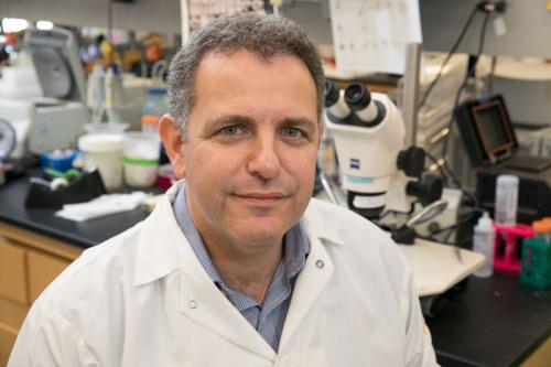
UCLA stem cell scientists go out on a limb for motor neuron disease
Often described as the final frontier of biology, the nervous system is a complex network comprised of the brain, spinal cord and the nerves that run through the body. Published today by scientists led by Bennett Novitch, Ph.D. at the Eli & Edythe Broad Center of Regenerative Medicine and Stem Cell Research at UCLA, new research using embryonic stem cells enhances the study of this intricate and perplexing system. Such discoveries are vital to the understanding and treatment of devastating disorders such as amyotrophic lateral sclerosis (commonly known as ALS or Lou Gehrig’s disease) and spinal muscular atrophy, which are caused by the loss or degeneration of motor neurons – the nerves that control muscle movement.
The research, which provides a more efficient way to study the functions of motor neuron diseases through the power of stem cells, was published online today by the journal Nature Communications.
Voluntary muscle movement—such as breathing, speaking, walking and fine motor skills—is controlled by several different subtypes of motor neurons. Originating in the spinal cord, motor neurons carry signals from the brain to muscles throughout the body. Loss of motor neuron function causes several neurological disorders for which treatment has been hindered by the inability to produce enough of the different kinds of motor neurons to accurately model and potentially treat neurodegenerative diseases, as well as spinal cord injuries. Specifically, efforts to produce ‘limb-innervating’ motor neurons in the lab were only about 3 percent effective. This special subtype of motor neuron supplies nerves to the arms and legs and is most acutely affected by motor neuron diseases such as ALS.
Novitch, an associate professor of neurobiology at UCLA and member of the Broad Stem Cell Research Center, studies the developmental mechanisms that drive muscle movements in order to better understand motor neuron diseases and discover new ways to treat them. Through the study, Novitch, the study’s first author Katrina Adams and their team sought to produce a better method to expand the range of motor neuron subtypes that could be created from stem cells, focusing on the subtype that innervates the limbs. Results from the study are impressive; the research found that the new method increased production of stem-cell-derived limb-innervating motor neurons from the previous 3 percent to 20 percent, more than a fivefold increase.
“Motor neuron diseases are complex and have no cure; currently we can only provide limited treatments that help patients with some symptoms,” said Novitch, the senior author on the study. “The results from our study present an effective approach for generating specific motor neuron populations from embryonic stem cells to enhance our understanding of motor neuron development and disease.”
Embryonic stem cells are ‘pluripotent’ which means they have the capability to turn into any other cell in the body. The process by which they turn into other cells in the body is called differentiation. Human embryonic stem cells are produced from stored frozen embryos donated by individuals or couples who have completed in vitro fertilization.

Using mouse and human embryonic stem cells, Novitch and his team directed the formation of limb-innervating motor neurons using a ‘transcriptional programming’ approach. This approach adds a transcription factor, which is a protein that acts in the cell nucleus to regulate gene function. Like a conductor guiding an orchestra, transcription factors tell the genes in a cell to turn off and on or express themselves a little louder or a little quieter.
Novitch’s previous research found that a transcription factor called Foxp1 is essential for the normal formation of limb-innervating motor neurons in a healthy spinal cord. However, most stem cell-derived motor neurons lack Foxp1, leading to the hypothesis that the addition of Foxp1 might convey limb-innervating properties. Novitch and his team tested this theory by first differentiating embryonic stem cells into motor neurons and then adding Foxp1. When transplanted into a developing chicken spinal cord, a widely used model for neurological research, the motor neurons modified with Foxp1 successfully grew out to innervate the limbs. This result, which is not usually seen with most unmodified stem-cell-derived motor neurons, illustrates the effectiveness of the Foxp1 transcription factor programming approach.
Scientists can now use this method to conduct more robust motor neuron studies that enhance the understanding of how these cells work and what happens when they deteriorate. The next steps in Novitch’s research include using the modified motor neurons to better understand how they are capable of innervating limbs and not other muscles, using similar transcriptional programming approaches to create other types of motor neurons and examining whether the motor neurons created might be effective in treating spinal cord injuries and neurodegenerative diseases.
This work was supported by grants from the California Institute for Regenerative Medicine, the Muscular Dystrophy Association, the National Institute of Neurological Disorders and Stroke and the Rose Hills Foundation, as well as support from the UCLA Broad Stem Cell Research Center.
