UCLA-led team induces cell reprogramming process with mechanical squeeze
UCLA bioengineers and colleagues have unveiled a fast, efficient and simple way to prime cells and help convert them from one type into another by squeezing them mechanically for a millisecond. This novel biophysical approach could improve the efficiency of cell engineering for tissue regeneration and the treatment of diseases such as cancer, Parkinson’s disease and muscular dystrophy.
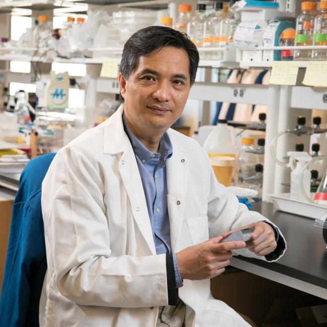
A research study on the advance, led by Song Li, Chancellor’s Professor of Bioengineering at UCLA, has been published in Nature Materials.
Cell reprogramming has been discovered and demonstrated since the early 1960s. It gives cells a new identity — imagine transforming skin cells into their embryonic stage, known as pluripotent stem cells, which can then be converted into mature cell types for the heart or muscles. The technique offers a promising way to treat a range of diseases by replacing damaged tissues with healthy ones. It could also open up new ways to model how diseases progress, test the effectiveness of potential drugs and make headway into personalizing treatments.
Unfortunately, the process of cell reprogramming has been largely laborious and inefficient. Similar to reprogramming a computer, to reprogram a cell, its memory needs to be reformatted. The biggest hurdle is the complexity of what’s known as the epigenome — the chemical “memory” made up of formatted DNA and DNA-affiliated proteins that dictates the cell type and how it functions.
Current techniques to induce cell reprogramming often involve using viruses to introduce genes into the nucleus that kick start the reprogramming process. Such techniques are inefficient, however, with success rates of less than 3% when converting adult skin cells into neurons, as an example.
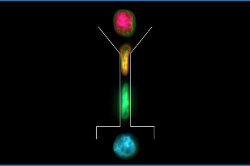
The UCLA team developed a non-chemical, biophysical approach to expedite that process by giving a cell nucleus a millisecond mechanical squeeze, which helped alter the physical and chemical traits of chromatin — a mixture of DNA and proteins that form the chromosomes found in the cells of humans and other higher organisms — and make them more prone to gene editing. The researchers developed a high-throughput microfluidic prototype with microchannels of 1/100th of a millimeter in diameter that squeezed cells as they moved through the device.
The near spherical cells were squished into oblong forms for a millisecond, and the slight deformation of the cell nucleus was just enough to partially open up the chromatin structure, thereby increasing accessibility to the genome.
The researchers found this method to be effective in priming cells for more efficient reprogramming, seeing 20% of cells reprogrammed from fibroblast cells into neurons. They also successfully applied the approach to various other cell types and reprogramming processes.
“This study explores the role of biophysical stimuli in reprogramming cells and decrypts some of the underlying mechanisms that influence that process,” said Li, who chairs the Bioengineering Department at the UCLA Samueli School of Engineering and holds a joint appointment at the David Geffen School of Medicine. “This provides a new pathway for improving cell reprogramming efficiency and in vitro cell engineering for regenerative medicine, disease modeling and drug screening.”
The researchers used a range of techniques from different disciplines to complete the research, including microfabrication, atomic force microscopy, fluorescence resonance energy transfer, nano mRNA sensors, epigenetic analysis and genetic engineering.
Authors of the study are UCLA researchers and graduate students Yang Song, Jennifer Soto, Binru Chen, Tyler Hoffman, Weikang Zhao and Chau Ly. The team also includes UCLA faculty members, Amy Rowat, a professor of integrative biology and physiology as well as bioengineering; and Dr. Siavash K. Kurdistani, chair and a professor of the Biological Chemistry Department. Other authors are Ninghao Zhu and Pak Kin Wong of Pennsylvania State University, as well as Qin Peng, Longwei Liu and Yingxiao Wang of UC San Diego.
The research was supported by a UCLA Eli and Edythe Broad Center of Regenerative Medicine and Stem Cell Research Innovation Award, and grants from the National Institutes of Health and the National Science Foundation. The researchers used the Nano & Pico Characterization Lab and Advanced Light Microscopy and Spectroscopy Lab at the California NanoSystems Institute at UCLA for part of the experiments.
The UCLA Technology Development Group has filed for a U.S. patent on the technology.
